Years of Pain and 50 Doctors Later..
Dental / Skeletal / Toxin Connection
This patient lives in Europe and flew in for a two-week dental visit.
Case Report: S.W. is a 35-year old female patient that presented with the following chronic symptoms:
- Head feels like it was unscrewed, and then put back on her shoulders again, but with such a twist that gives the feeling that the upper face has gone to one side and the lower face to the other side. The word ‘TORN’ would describe it best.
- Pain in left side muscles from top of head to toes.
- Nausea.
- Hole where my lower right first molar was extracted is still not completely closed and pain occurs with pressure on area.
- Lower jaw feels like it has to be moved to the right.
- Dry eyes; dry membrane syndrome relating to women’s problem.
- Left side muscle pain at the top of my neck.
- 8 years of overall pain.
- Constant pain for past two years in the upper left first molar; also pain between this tooth and the one behind it; gum in this area bleeds.
- Right side of my bite feels like it needs to be built-up. Needs more defined cusps.
- Bloated stomach.
- Worst pain is in the right and left side of my neck and cranium.
Cranial Evaluation: Revealed that the patient’s head was literally lock up without much pumping motion for movement of the cerebrospinal fluid. Also noted was the fact that when the patient closed her teeth together, the skull distortions worsened. This finding establishes the fact that the patient’s bite is off and treatment must focus on correcting the teeth, i.e., rebuilding height on some and modifying the contours on others. Since the teeth function like gears, they must be made to mesh evenly at the correct vertical height to balance the skull bones, spine and pelvis.
Dental Findings: The patient recently had two transitional gold and resin crowns fabricated. Unfortunately, the patient was allergic to the gold that was used in these two crowns. In addition, a previously root canalled tooth (upper left first molar) had a mercury filling left in the tooth as well as having a residual infection in the root structures. Further more the temporary crown placed on this molar tooth had a poor anatomical form and no contact with the adjacent tooth which permitted food impaction and ulceration of the gum. Two major muscle groups that attached to the lower jaw were in severe spasm. Also of interest was the patient’s sensitivity to mercury, gold, cobalt and chromium as well as gluten. There was a neurologic interference on the upper left first bicuspid tooth that helped trigger off the muscle spasm.
Eighteen months prior to the patient’s visit, the lower right first molar was extracted. Although the x-ray of the area looks totally normal, an Osteomyelitis (inflammation of the bone) was present in the socket where the tooth was removed. Also present were a group of pathogens in some of the teeth and jaw bone (Streptococcus, Pseudomonas, and Mycoplasma.)
Subluxations: There were numerous joints in the body with misalignments of the anatomical parts: Left shoulder, left medial pole of elbow, left foot (middle two toes), right leg lateral knee, right foot (4th digit) and right hand 3rd digit. These misaligned joints provided a constant barrage of faulty impulses into the nervous system and directly caused structural imbalances of the spine, jaw and skull. The patient also presented with a Category II (weak ligaments) right sacroiliac weakness.
Neurologic muscle testing revealed functional weakness on the patient’s left side: Cervical vertebrae C2 to C6 and the left pelvic muscles. The upper left trapezius muscle (where the shoulder meets the neck) was very painful to palpation. There was a bite interference on the lower right second molar that was the direct cause for all left-sided muscle weakness.
Oral pH: 4.5 (Normal 6.8 to 7.2) The extremely low acid level has the clinical significance of drastically lowering the patient’s pain threshold. The slightest imbalance will be perceived as extremely painful. Also the more acidic the tissues the greater the reactivity of heavy metals (mercury, gold, cobalt, chromium).
Thyroid Dysfunction: The patient exhibited an under-active thyroid, which was directly caused by mercury in the gland. The lymphatic system (sewage system) that drains the lower jaw goes directly to the thyroid gland carrying heavy metals and other substances (bacteria, viruses, etc.). The key to resolving this problem is removal of the mercury from the thyroid tissue with chelating nutrients.
Metabolic Status: The patient tested out to be a Fast Oxidizer and Parasympathetic Dominant. Being a fast oxidizer means that the individual metabolizes or burns their food rapidly. Diet wise these type patients must avoid eating refined carbohydrates (white rice, white bread, white sugar, etc.) and consume a dietary regime that approximates 40% protein, 30% fats and 30% complex carbohydrates. These foods function to slow down the fast oxidation process and shift the parasympathetic dominance toward sympathetic function. Once balanced a broader diet can then be consumed.
Impression: The patient presents with a multitude of factors that are contributing to her chronic pain. Her extremely low oral pH of 4.5 signifies loss of alkaline reserves (minerals: Ca, Mg, K) and an extremely low threshold to pain, which is clinically obvious. Also present is an acutely sensitive occlusal awareness (slight changes in biting pressure cause an over reaction of the nervous system). Two major dental foci are present: the infected root canalled upper left first molar and post extraction space of the lower right first molar. The patient’s metabolic typing (fast oxidizer/parasympathetic dominant) is such that consumption of carbohydrates will worsen her condition by lowering the pH, destabilize blood sugar and heighten an allergic reaction with gluten. The cranial distortions and poor occlusal relationship are destabilizing her upper cervical vertebra, spine, pelvis and cranium. There is a fascial restriction over the spleen/stomach/pancreas area, which I believe is compensatory to the dental foci (infected upper left first molar) and bit interferences. The subluxations listed provide a constant barrage of faulty signals into the nervous system, which in turn contribute, to a distorted body posture. An additional dental foci is the presence of the allergy to the gold in crowns #12 & 14. Because of the multitude of factors, it is my opinion that removal of the toxic mercury fillings should only be accomplished after the patient has had a time to detoxify and correct the metabolic imbalance.
Biopsy Report: Confirmed the presence of Osteomyelitis in the lower right extraction space (where first molar was removed 18 months ago).
Treatment – Phase I
- Remove gold crowns #12 & 14 and replace with provisional crowns. Done.
- Remove mercury filling in #14. Done.
- Fabricate new provisional crown for #14 correcting bite and distal contact. Done.
- Cavitational surgery to remove infected bone #30 and fill defect on mesial of #31 with PerioGlass (synthetic bone). Done.
- Cranial manipulation coupled with occlusal adjustment/build-ups to help stabilize the cranium. Done but needs follow-up treatment.
- Adjust subluxations as described in the report. Done but needs follow-up treatment.
- Correct neurologic occlusal interferences. Done
- Dietary correction of metabolic typing Fast Oxidizer/Parasympathetic Dominance. In the process.
- Detoxification of heavy metals. Just started.
The patient was nutritionally evaluated and placed on a custom supplement program and Group II diet.
Progress – Major changes that occurred during the two-week visit included:
- 100% reduction of pain in the upper left first molar tooth that was constant for a two-year period.
- 95% reduction in left side muscle pain.
- 30% reduction in feeling of the head not being properly positioned on the neck.
- 40% reduction in left side muscle pain from top of head to toes.
- 50% reduction in stomach bloating.
- 70% reduction in eye dryness.
- 60% reduction in nausea.
Changes that were reported within two weeks of returning home to Europe:
- 80% reduction in menstrual cramping.
- 60% increase in overall energy and well being.
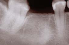
Lower right first molar was extracted in September, 2000. The extraction socket was left exposed to the oral fluids. In addition the periodontal membranes were not curetted out and the infected bone was not removed. The condensing osteitis was the result of mercury leaking down the root into the surrounding bone. Cavitational surgery was performed on 3/8/02 and the patient’s left cervical pain of 18 months duration decreased by 95% when the anesthesia wore off.
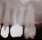
The upper left first molar had an infection in the dentine tubules. Molars have approximately 3 miles of dentine tubules which provide refuge for anaerobic pathogens. There was a mercury filling in the palatal canal from the access opening. Direct resonance testing diagnosed the presence of streptococcus viridans, pseudomonas, mycoplasma, osteomyelitis and gangrenous pulp. The patient was also allergic to the gold in the cast metal/acrylic crown. Furthermore the distal contact was open and allowed food to trap causing the interproximal gingiva to ulcerate. This tooth provided constant pain for two years. Appropriate treatment resolved the pain in just three days. Tx: Hg removed, pulp chamber cleansed and medicated with lavender oil, gold/acrylic crown was cut off and a new temporary fabricated to correct compatibility and distal contact. Bite was adjusted neurologically. Pain of two years duration resolved in three days.
Eighteen months prior to the patient’s visit, the lower right first molar was extracted. Although the x-ray of the area looks totally normal, an Osteomyelitis (inflammation of the bone) was present in the socket where the tooth was removed. Also present were a group of pathogens in some of the teeth and jaw bone (Streptococcus, Pseudomonas, and Mycoplasma).
FREE PRESENTATION
Download the slides from Dr. Smith's presentation on the Dental Whole Body Connection in Ontario on October 25, 2024.
A comprehensive seminar to awaken you to the potential dangers of dental treatments.

STAY INFORMED
Big tech and mainstream media try to suppress the powerful information I have to share. Subscribe here to stay informed!