Bruxism: Dental/Cranial Connection
J.S. bruxed or ground her teeth on a regular basis. The traditional concept is that bruxism is due to emotional stress. Patients who present a chief complaint of bruxism to their dentist are most often offered two treatment approaches: The first approach focuses on “removing the tooth interferences” which cause the muscles to tighten up. This treatment is referred to as an occlusal adjustment. Dentists use articulating paper (red and blue carbon impregnated paper) to mark the “high spots” or tooth interferences and them judiciously grind them off. The second approach uses a night appliance to protect their teeth while the patient grinds their teeth during sleep. Both treatments are within the dental standard of care. Both treatments have attributes and have helped many patients. In a percentage of cases, removing a “high spot” or working interference makes the patient’s bite more comfortable and reduces the bruxism. The appliance on the other hand focuses on protecting the teeth while the patient grinds their teeth during sleep. Although beneficial use of the appliance does not solve the underlying problem.
J.S. underwent routine dental treatment involving two teeth which required root canal therapy. Her problems started when both treated teeth were capped with gold and porcelain crowns. The bite on these crowns was not comfortable. The treating dentist proceeded to adjust her bite by grind off interference points on the two crowns but also on several posterior or molar teeth. This procedure was performed at least three times and after each treatment the patient experienced more neck and facial pain. This process started in the year 2000 and continued to march of 2005. During those five painful years, The patient had her teeth capped with permanent crowns. Because they were so painful and could not tolerate the bite, they had to be removed and temporary crowns placed. In addition, the patient had to drop out of graduate school because the pain prevented her from concentrating and retaining information. Dental treatment during that time included capping a number of posterior teeth and no less than ten TMJ appliances. Referrals to various TMJ and oral surgeons brought no relief. The last recommended treatment offered was to irrigate her jaw joints to “flush out” possible debris. Fortunately the patient did not see the merit in this recommendation.
Evaluation of J.S. showed a severe cranial bone misalignment. The bones of the skull are joined together by means of sutures, which act as expansion/contraction joints. The teeth function as the self-correcting mechanism to realign the skull. This mechanism operates through the maxillae or “upper jaw.” The maxillae represents the anterior two-thirds of the base of the human skull. When the teeth are properly aligned contact during normal swallowing and chewing “resets” the alignment of the maxillae, which in turn rebalances the rest of the skull. When patients have a malocclusion or crooked teeth, a distortion occurs to the alignment of the the skull. The misalignment of the skull in turn causes an increase in tension of the fibrous membranes that act as internal tent poles to support the skull. These fibrous membranes are innervated with sensory nerves which trigger off pain. This membrane surrounds the brain and continues through the base of the skull (foramen magnum) and attaches to the neck vertebrae and continues all the way down to the tail bone (coccyx). In essence, the skull, spine and pelvis function like a slinky. A distortion at one end will cause an equal and opposite distortion somewhere along this membrane tube. This craniosacral system is the reason why patients experience symptoms far removed from the actual site of the original problem. When dentists grind down the supporting enamel on molar and bicuspid teeth they cause the spine to be compressed. This was the case with JS. Every time the dentist ground down her back teeth he compressed her spine more and her pain increased. In addition, removing back molar support now allowed her lower front teeth to hit into her upper front teeth. Collision of the upper and lower front teeth cause the neck flexors (muscles on the side of the neck) to go into spasm and pain. J.S. had to resort to pain medication and sleeping pills. March 15, 2005 was the first time in many years that J.S. did not have to take any medication. On March 14, her posterior teeth were built up to level her bite. The discrepancy was the thickness of three sheets of typing paper on the left side and two on the right. Dentists cannot make an appliance this thin. Every appliance that was made just served to increase the distortions in the skull because they did not correct the high cant of the horizontal plane. Since the foundation of the skull was never corrected, the patient’s problem was never addressed. J.S. suffered five years because the treating dentists failed to recognize this distortion. Every skilled craftsmen except dentists know that the foundation must be leveled before any structure can be properly built. The clinical photos clearly exhibit the structural distortion and have been provided for readers to comprehend the dental structural dynamics.
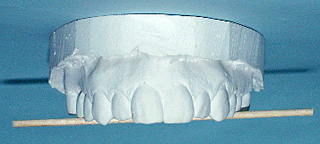
The horizontal plane cants upward on the left side. This was worsened by dentists grinding down the patient’s teeth to remove biting interferences. Every time the patient closed her mouth the lower jaw closed higher on the left side than the right. Since there are 136 muscles that are directly affected by the lower jaw, one can readily appreciate the domino effect that is brought into play.
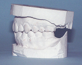
This left side view shows the elevated height of the molar and bicuspid teeth.
The lower right posterior teeth were also grown down contributing to the compression of the spine and allowing the lower front teeth to hit into the upper incisors.
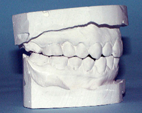
The upper left posterior teeth were built up by means of bonding dental resin material directly on the teeth. Adding the thickness of only three sheets of paper was sufficient to rebalance the skull bone alignment. In essence it is like shimming up a window or door frame. In this case the skull is being shimmed.

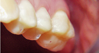
The lower right posterior teeth were also built up with dental resins by only two thicknesses of typing paper. Clinically the exact height is determined by using four cranial indicators. By “shimming up the teeth a skilled practitioner can immediately tell when the skull bones are in perfect alignment. Dr. Smith is the originator of using specific cranial indicators to assess cranial bone alignment and the new shimming technology which has been used to successfully treat many chronic pain patients.
It has been this researchers experience that a high percentage of patients who brux their teeth do so as a reflex mechanism to wear away tooth interferences and force the skiull bones into a better alignment. This observation is based on the fact that when patients’ bites and skulls are aligned at the same visit they report back that their need to brux or gind their teeth greatly diminishes or totally disappears.
… when patients’ bites and skulls are aligned at the same visit they report back that their need to brux or gind their teeth greatly deminishes or totally disappears.
FREE PRESENTATION
Download the slides from Dr. Smith's presentation on the Dental Whole Body Connection in Ontario on October 25, 2024.
A comprehensive seminar to awaken you to the potential dangers of dental treatments.

STAY INFORMED
Big tech and mainstream media try to suppress the powerful information I have to share. Subscribe here to stay informed!