Dental / Migraine Connection
Conventional medicine still does not know what causes migraine headaches. Various theories exist which focus on blood flow irregularities (dilation of blood vessels), psychological and chemical changes (hormonal, toxicity and heavy metals, etc.) and triggers (bright light, smells, foods, low blood sugar, weather changes, aspartame and other additives). One factor often overlooked by the medical establishment is structural distortions stemming from cranial dental imbalances.
Many of the symptoms plaguing migraine patients have their origin from stimulation of the autonomic nervous system (ANS). The ANS primarily deals with the peripheral parts of the body as opposed to the central nervous system (CNS), which monitors the brain and spinal cord transmissions. The ANS works like the gas pedal (acceleration: sympathetic) and braking (slow down: parasympathetic) systems of your car. An increased heart rate results from activation of the sympathetic nervous system. Eating stimulates the parasympathetic system, which works to aid digestion by stimulating release of digestive enzymes. The two components work to counter balance each other. The parasympathetic nervous system monitors the skull and pelvic regions where as the sympathetics deals with the thoracolumbar part of the spine. If the cranial bones and sutures (expansion and contraction joints between skull bones) are distorted then changes in pressure from weather fronts or ascending and descending in an airplane can trigger off nausea, vomiting, diarrhea and migraine pain.
The key to diagnosing dental cranial distortions lies in the use of four indicators to palpate the skull and determine whether or not teeth contact worsen the cranial strain patterns. If teeth contact does increase the existing skull distortion, then a high probability exists that this factor is a major cause for migraine headaches.
K.T. was only fourteen years old and had experienced migraine headaches for the past ten years. She had undergone orthodontic treatment once, which did not alter the headaches and the straightened teeth relapsed within a few years. A craniopath referred Kate to our office. Examination determined the presence of cranial distortions with a worsening when she closed her teeth. Treatment involved use of the ALF (Advanced Lightwire Functional) appliance to correct the cranial torsions or strain patterns in the skull. Once the skull bone alignment was completed, conventional braces were used to align the teeth to hold the cranial bone correction. Treatment took two years and Kate has been totally migraine free since correction was achieved. The advantage of a structural correction is the use of a non-invasive procedure, plus the underlying cause of the problem is fixed.
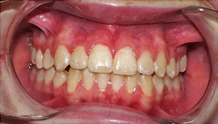
Figure 1.
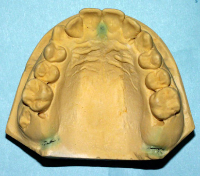
Pre-treatment: Maxillary or upper arch exhibits a lack of arch length, which resulted in right side crowing. This dental deformity represents more than just a physical space discrepancy. Crowding is accompanied by cranial bond distortions, which have tension patterns within the dural membrane system. It is the tension, which causes stimulation of the nerves, which triggers off the migraine headaches. By expanding the jaw bone and removing the tension pattern resolves the migraine headaches.
Figure 2.
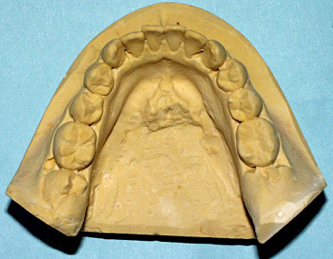
Pre-treatment: Lower dental arch exhibits minor crowding in the front and posterior teeth and arch length discrepancy mimicking the upper right side.
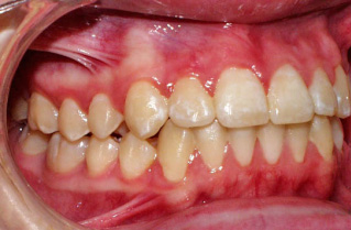
Figure 3.
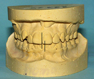
Pre-treatment: The maxillae or upper jaw is slightly rotated to the right. In addition, there is a slight loss of vertical height due to failure of the posterior teeth to fully erupt. Another major factor in migraine headaches is the abnormal tensions generated from teeth contact. Malocclusion or crooked teeth and jaw alignment have the potential of creating abnormal tension within the dural membranes in the skull. Very few dentists are knowledgeable or have the skills to diagnose cranial distortions generated from malocclusion.
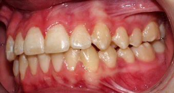
Figure 4.
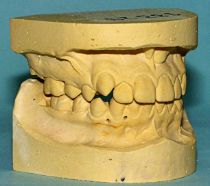
Pre-treatment: The arch length shortage on the right side is responsible for the crowding of the upper canine tooth.
Figure 5.
Post-treatment: Not only was the mid-lines of the upper and lower teeth and dental arches corrected but the posterior teeth were erupted and vertical dimension or jaw height corrected. In addition, the cranial distortions, dural membrane tension resolved. The ultimate goal however is to align the teeth with braces after the cranial bones are re-aligned so that when the teeth come together as in chewing or swallowing the cranial bones remain aligned and the dural membrane system remains relatively tension free. In the final analysis it is the achievement of this last phase of treatment that is the most important. Ninety-nine and nine-tenths percent of all dentists have no clue or knowledge that this crucial concept even exists. Most orthodontists jiggle teeth around to make them straight but they do not correct the cranial base in which the teeth are imbedded. This I believe is one of the reasons why there are so many structural problems (chronic neck, arm, hand, low back, knee, and ankle problems). And because very few dentists or even chiropractors have the diagnostic skills the patients problems linger indefinitely.
Figure 6.
Post-treatment: The arch length discrepancies of both arches were corrected by literally stretching the bone with the ALF Functional appliances. The correction process is more than just aligning teeth.
Figure 7.
Post-treatment: This patient is migraine free after suffering for ten years. The key is to balance the teeth, cranial bones and dural membrane systems. No amount of drugs, manipulation or acupuncture can correct a structural problem. Clinically, diagnosing or defining the underlying problem is the only way to achieve a cure.
Figure 8.
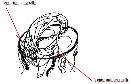
Post-treatment: The abnormal tensions within the dural membrane system must be removed in order to resolve the migraine pain. The reason half the head is in pain is because one half is in flexion (relative tension) while the other half is in extension (relative relaxation). All the membranes above and including the tentorium cerebelli are innervated by the three divisions of the fifth cranial nerve (trigeminal nerve).
Many of the symptoms plaguing migraine patients have their origin from stimulation of the autonomic nervous system (ANS).
FREE PRESENTATION
Download the slides from Dr. Smith's presentation on the Dental Whole Body Connection in Ontario on October 25, 2024.
A comprehensive seminar to awaken you to the potential dangers of dental treatments.

STAY INFORMED
Big tech and mainstream media try to suppress the powerful information I have to share. Subscribe here to stay informed!