Dental / Head-Face-Neck-Shoulder Pain Connection
A 16 year-old male hit the left side of his head on a basketball pole while going up for a lay-up shot. Even though the pole was padded the impact was sufficient to cause a concussion and the patient was knocked unconscious for 3 minutes. As a result of the head trauma, the patient suffered severe left sided symptoms: constant headaches, retrobulbar (behind the eye) eye pain, tinnitus, facial pain in the region of the cheek, shooting neck and shoulder pain. The patient also had hyperacusis (normal sounds perceived as loud), insomnia and dizziness. Extensive medical work ups by neurologists, ENT, internists and psychiatrists produced no definitive diagnosis. Various medications were prescribed with no resolution of symptoms. These symptoms were present for a seven month period and prevented the patient from attending school.
Cranial Evaluation
The impact had caused a reversal of the normal pumping motion of the skull bones, decreased expansion on the left side of the cranium, left internal rotation (compression) of the temporal and malar bones, high right sphenoid with torsion in relationship to the occiput (bone that makes up the back of the skull).
Dental Evaluation
The patient had a mild skeletal discrepancy in which the lower jaw was slightly larger than the upper. He had a misalignment of the bite on the right posterior section of teeth. This malocclusion existed prior to the incident but was accompanied without any symptoms. Extensive muscle spasms were present. They involved the right and left external pterygoids, which attach from the neck of the jawbone where it forms the TM joint to a bony plate of the sphenoid bone. The sphenoid bone is very important since it houses the pituitary gland (master endocrine gland) and has an extensive dural membrane attachment. These structures are responsible for many of the undiagnosed headaches, ear, neck and facial pain.
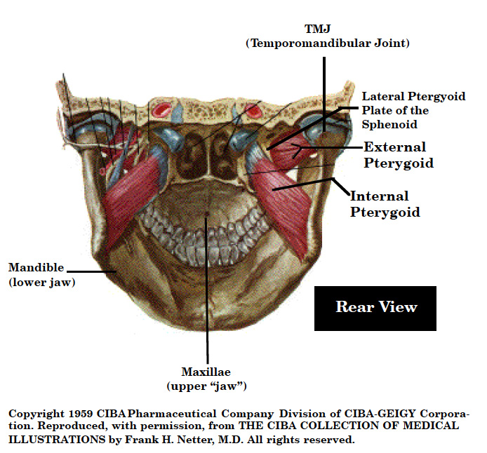
The internal and external pterygoid muscles act as a sling system for balancing the sphenoid bone. Whenever there is an imbalance of the teeth cranium, or upper cervical vertebrae, these muscles, will have a great propensity for going into spasm. The constant pull of these imbalanced muscles places tension on the sphenoid bone which affects the pituitary gland (master endocrine gland), dural membrane attachment (all the way down to the sacrum), extensive cranial motor and sensory nerves, all three major divisions of the trigeminal nerve, blood supply to the dural membrane system, tensor tympani muscle to the ear, muscles of the eye and places a drag on the pumping mechanism for the cerebrospinal fluid. These distortions are responsible for numerous neurologic symptoms: headaches, eye pain and visual distortions, conductive hearing loss, tinnitus, hyperacusis, equilibrium problems, memory problems, facial pain, fatigue, low blood sugar, numbness and pain in the neck, shoulder and arms.
Treatment Sequencing
- Cranial manipulation was performed to correct the reversed cranial motion and free up the compressed skull bones.
- Micro-current stimulation combined with myofascial release techniques were used to release the spastic muscles.
- ALF upper and lower appliances to correct cranial stress patterns.
- Glutathione was used to detoxify the patient’s liver from all the medications that were used.
Prognosis: Excellent
The hyperacusis or loud sounds were resolved and the headaches greatly reduced following the third cranial adjustment. Following two months of treatment with ALF appliances the cervical and shoulder symptoms and insomnia cleared. The appliances were discontinued following resolution of symptoms.
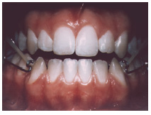
Figure 1. The ALF appliances are used to gently expand the upper arch (maxillae) to free up the cranial bones and reduce the internal stress pattern.
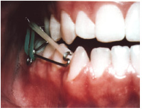
Figure 2. The vertical green elastic is used to bring down a high sphenoid bone while the tan elastic is being used to help bring the maxillae forward.
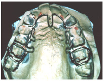
Figure 3. The upper ALF appliance is tacked onto the front teeth by means of light cured resins. By so doing the entire maxillae is treated as a single unit. There is no plastic on these appliances which increases their comfort and does not impair speech.
Use of the ALF appliances affords the dentist a non-invasive means of correcting structural strain patterns which in turn have the potential to cause neurologic and physiologic changes throughout the body. No other medical specialty has the technology to make these types of changes. Use of the ALF appliance complements the osteopath, chiropractor, physical therapist, psychiatrist, orthopedist and neurologist as well as the podiatrist.
Patient suffered severe left sided symptoms: constant headaches, retrobulbar (behind the eye) eye pain, tinnitus, facial pain in the region of the cheek, shooting neck and shoulder pain.

STAY INFORMED
Big tech and mainstream media try to suppress the powerful information I have to share. Subscribe here to stay informed!