Dental Migraine/PMS/Low Back and Neck Pain Connection
Patients who have had four bicuspid teeth removed for braces bear structural distortions throughout life. Since most orthodontic treatment takes place during the teenage years, the symptoms that develop are often attributed to social pressures or hormonal imbalances. Unfortunately the real cause for the symptoms is not recognized and the patient is left to suffer. A case in point was Yvette D.
Yvette was a 44 year-old white female who 25 years earlier had four first bicuspid teeth amputated to make room to “straighten” the remaining teeth. At the time that she had her teeth surgically removed and the conventional braces placed, she developed migraine headaches. During the twenty-five years of being plagued with migraines she also developed PMS, low back pain, left neck and shoulder pain. The left neck and shoulder pain was also accompanied by a severe pulling as if a cord was constantly being stretched. Needless to say medical treatments, drugs nor chiropractic manipulation had any lasting affect on the cluster of symptoms.
Yvette’s case involved an overlay of five factors which prevented resolution by conventional treatment. First, there was the cranial distortions caused by the extractions. Second, a deep overbite existed. Third, the planes (foundation) of her maxillae were not level. Fourth, there was psychological distress in her work place. And finally, there were nutritional deficiencies due to a diet high in processed foods. The proposed treatment plan involved a strategy to correct the underlying factors:
- Factors 1 & 2: Overlay resins were constructed to support the collapsed vertical and also level the maxillae (foundation).
- Factor 3: ALF appliances to correct the cranial distortions.
- Factor 4: Psychological counseling to resolve the distress issues at work.
- Factor 5: Nutritional support to correct existing deficiencies.
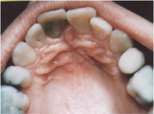
Figure 1. Amputation of the first bicuspid teeth caused the maxillary (upper) arch to collapse as seen in the “V” shaped narrowed and distorted form. If extracting teeth was the correct solution, then adequate space should have provided a stable situation and allowed proper alignment of teeth to remain. Removing the teeth worsened the situation by causing the muscles around the mouth to tighten and cause relapse. The pre-maxillae, bone that holds the upper four front teeth, is now jammed and restricts the fourteen facial bones. These restrictions set up stress patterns in the membrane system and skull bones that ultimately affects the neck, shoulder and low back.
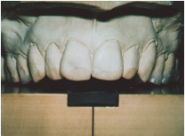
Figure 2. The upper teeth are being analyzed on the Accu-Liner instrument. Those teeth that do not touch the horizontal stage must be built-up to even the pressures to the skull bones. The technique is analogous to leveling the ground prior to laying the foundation of a house.
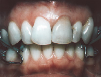
Figure 3. The ALF appliances are in place. The green orthodontic elastics are being used to correct existing torsions, strain patterns and to move the entire maxillae forward.
During the three years of treatment the strain patterns were gradually taken out of the skull, the tilts of the maxillae were improved, and the stress patterns in the dural membrane system that surrounds the brain were released. The patient was last seen in October 1997 and has been completely free of migraines, neck, shoulder, low back pain and PMS symptoms for the past two years.
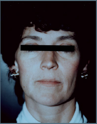
Figure 4a. Pre treatment: note tilt of the patient’s mouth. It is high on the patient’s right side. Also note the fullness of the face which occurred as a result of expanding the maxillae (base of the skull) closer to its original size.
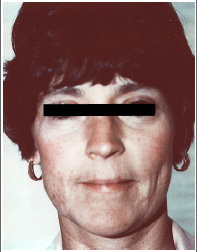
Figure 4b. Post treatment: note the dramatic change on the level of the mouth. Correcting the foundation (maxillae-upper jaw) levels the foundation of the skull, dural membrane system, neck vertebrae and all the associated muscles.
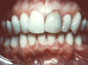
Figure 5. Completed case: The dental arches have been corrected by widening the front portion of the arches and the teeth aligned to stabilize the skull bones. With extraction of four bicuspid teeth during the teenage years, the genetic potential for complete facial growth is diminished and total correction of the skull bone distortions is virtually impossible. Even though the distortions could not be totally corrected the pain was totally resolved because the system was put back into a functional range which the body could handle.
Patients who have had four bicuspid teeth removed for braces bear structural distortions throughout life.

STAY INFORMED
Big tech and mainstream media try to suppress the powerful information I have to share. Subscribe here to stay informed!