Pitfalls of Third Molar Extraction
Kala T. had her wisdom teeth removed in 2011 and five months post-extraction her whole head started to change shape. Her forehead started to push outward, back of her head as well as all the bones started to bulge outward. Following the skull bone changes her shoulders, hips and down to her feet started to shift position.
This process continued for approximately two years. Numerous specialists were consulted with no positive outcome.
Cranial evaluation revealed specific cranial distortions were present. One of the key issues was the fact that removal of her four third molars caused her maxillae to constrict, which in turn forced the remaining bones to bulge outward. Third molars or wisdom teeth provide essential support to the cranial vault. Extraction may result in loss of cranial bone alignment, which 99.99% of dentists have no knowledge.
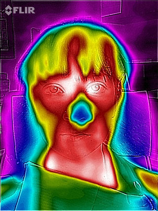
Treatment involved placement of an ALF maxillary ultra-light appliance. Within six weeks of initial expansion to correct the specific cranial bone distortions, Kala stated that her skull started to feel more normal. During the fourth visit, Digital Infrared Thermal Imaging was used to document a baseline vasomotor status and cranial bone shape. The pre-adjustment DITI image exhibits severe restriction of blood flow to the mid-face and nose, two vertical projections of increased blood flow to the frontal lobe, misshaped cranial vault and highly inflamed muscle attachments at the clavicular and nasal areas.
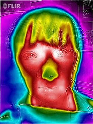
The patient’s cranial indicators were noted in her chart, the ALF was then removed and adjusted to correct the specific cranial distortions noted. The ALF was then replaced in the patient’s mouth. A five minute wait period was allowed to permit structural and vasomotor changes to take place. The post adjustment DITI image was taken and shows dramatic changes: blood flow to the mid-face and nose dramatically improved; the shape of the cranial vault became much more symmetrical; vertical blood flow to the frontal lobe evened out and the previously inflamed clavicular area greatly diminished. The patient stated that her head felt much more balanced and normal. Use of the cranial indicators to guide appliance correction is now accomplished by design rather than taking an unscientific approach to just expanding. The Advanced Light Wire Functional (ALF) appliances represent the biggest breakthrough in orthodontic technology since its inception 175 years ago.

STAY INFORMED
Big tech and mainstream media try to suppress the powerful information I have to share. Subscribe here to stay informed!